Search
"Crazy coworker stabbed her at her house." (Liver laceration with active bleeding, hemopneumothorax.)


41 year old female who was stabbed through her right inframammary fold (aka under her right breast).
CT through 3 sequential phases shows a linear laceration through the right lobe of the liver. There is a small focus of contrast extravasation that enlarges over time, eventually spilling out of the liver into the peritoneal cavity. These findings are consistent with an AAST grade IV liver injury.
Oh, the knife also apparently went through the liver and diaphragm, into the chest cavity, because she also had a hemopneumothorax.
Her blood counts were trended and were stable, so she didn't need surgery for the liver laceration. She did have a chest tube placed for the pneumothorax.
Acute alcoholic hepatitis.


50 year old female with history of alcohol abuse.
Baseline (6 months before): CT shows diffuse moderately low attenuation liver compatible with (alcoholic) fatty liver disease.
Patient was admitted for septic shock. CT shows a markedly enlarged liver (compare the left margin of the liver [red arrows]), markedly decreased liver attenuation, and engorged portal veins.
Subsequent CTs show the diffusely low liver evolving into a heterogeneous, splotchy, appearance and development of edema [green arrows] from decompensated liver failure. Her total bilirubin rose from baseline 0.4 to 16.4 at the 2 week mark.
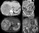
Another cholangiocarcinoma.


68 year old with several months of worsening fatigue and 20 pound weight loss. Physical exam notable for a firm, nodular liver.
CT [top images]: Large round, heterogeneous mass, this time in the right hepatic lobe. See here for a left hepatic cholangio. This was initially thought to be a large hemangioma.
MR [bottom left image - T2 FS, bottom right image - postcontrast LAVA]: Large, very heterogeneous mass, partially compressing the inferior vena cava.
This mass was also not survivable.
Cholangiocarcinoma and choledochocele.


59 year old with 6 months of abdominal pain and weight loss. CA 19-9 tumor marker measured 92,000. CEA tumor marker measured 205.
CT [top images]: Large heterogeneous mass with irregular borders involving the left hepatic lobe.
CT [bottom images]: Protrusion of the distal common bile duct (red arrow) into the duodenum, likely represent a Todani type III bile cyst / choledochocele.
This aggressive cancer was not survivable.

