Search
Life's middle finger: malignant melanoma.


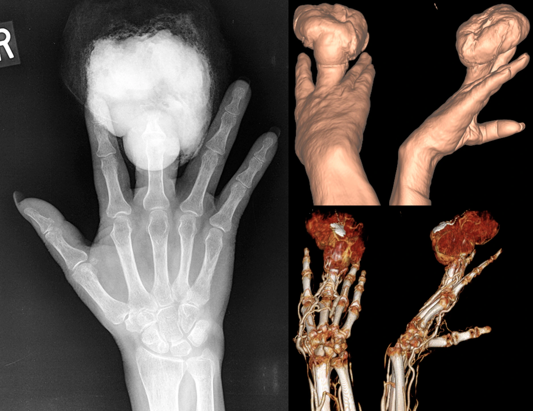
54 year old female with a history of minor injury to the finger 6 months before these studies were obtained, subsequently developed an infection requiring debridement. The wound was then stable until 2 months before, when a large, fungating, hemorrhagic mass grew.
X-ray shows large radiopaque mass eroding the distal finger (distal phalanx and distal portion of the middle phalanx).
CT 3D surface reconstructions show the morphology of the mass.
CTA 3D reconstruction shows the mass is very hypervascular.
Lymph node scintigraphy was performed showing a sentinel node at the axilla (not shown). The patient underwent amputation of the finger with axillary sentinel node biopsy, which was positive for metastatic melanoma. The patient was then lost to follow-up.
Leriche syndrome (aortoiliac occlusive disease).


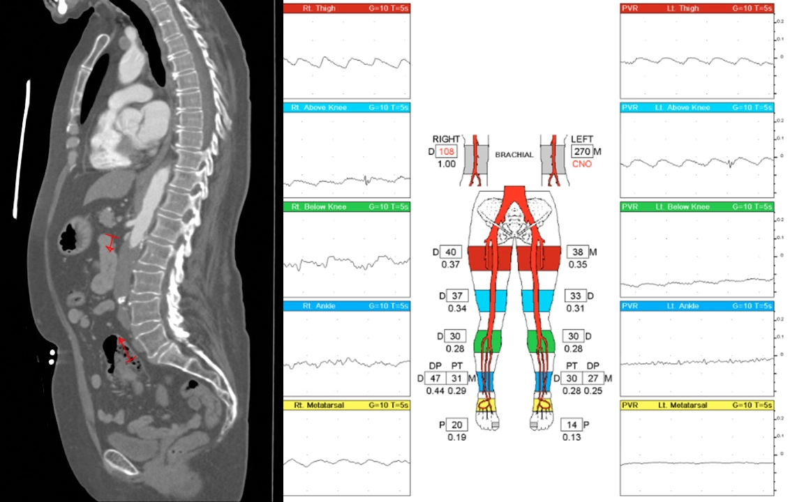
62 year old female with a history of tobacco, opioid, and meth abuse as well as heart attack requiring coronary bypass and stents. She presents with bilateral lower extremity claudication (painful fatigue with physical activity) starting from her glutes.
CT angiogram shows occlusion of the lower aorta (red markings) as well as both common iliac arteries.
Vascular ultrasound shows very poor arterial waveforms throughout the lower extremities, essentially flat/absent below the knee.
She underwent a transabdominal aortobifemoral bypass to treat this.
Radiopedia article on Leriche syndrome.
Lethal left ventricular rupture.


Elderly female with a history of hypertension, stroke, and, 1 month before, heart attack with cardiac arrest. The patient presented to the emergency department with 1 day of nausea, vomiting, cold sweats, and malaise. Troponins 1.54. ECG with inferior Q waves but no ST elevation/depression or T-wave inversion.
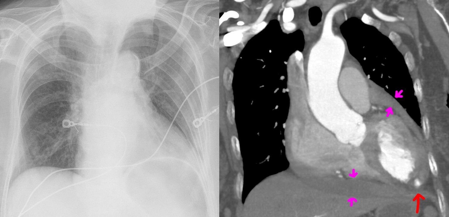
Chest radiograph shows an enlarged cardiac silhouette, for which a differential of cardiomegaly or pericardial effusion was suggested, as well as increased pulmonary vascular markings compatible with pulmonary congestion. Compared chest radiograph findings to the images from the methamphetamine-induced cardiomyopathy case.
CT angiography shows both cardiomegaly as well as an intermediate-density pericardial effusion (magenta arrows) concerning for hemopericardium. Most concerningly (holy-shit concerningly), there is a left ventricular aneurysm with rupture and blush of contrast (red arrow) into the hemopericardium, compatible with ongoing bleeding.
It is not surprising that a rupture of the heart carries high mortality risk. The patient passed away from PEA cardiac arrest shortly after admission, likely from cardiac tamponade by the enlarging hemopericardium.