Search
Grynfeltt-Lesshaft hernia. (This will be the last case I post for a few days.)


Incidental finding of a superior lumbar hernia (Grynfeltt-Lesshaft hernia). In this case, only a lobule of retroperitoneal fat is herniating through the defect, but organs can also herniate through.
Inguinal hernia containing bladder.

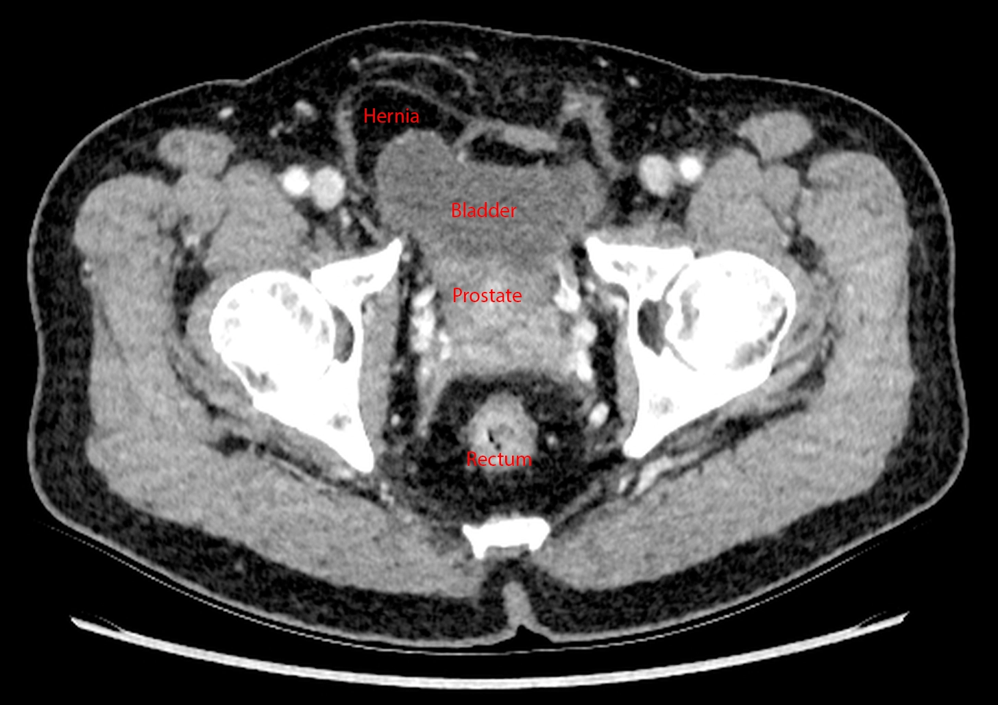
Continuing the theme of things extending into spaces they don't belong in, this is an incidental finding of an inguinal hernia that contains a small portion of the bladder. The patient got the CT for other reasons.
Bowel into inguinal hernia causing bowel obstruction.
Appendix into inguinal hernia, incidental finding.
Ventriculoperitoneal shunt into inguinal hernia, incidental finding.
Abdominal distention despite being told her uterine fibroids "shrunk" after menopause.

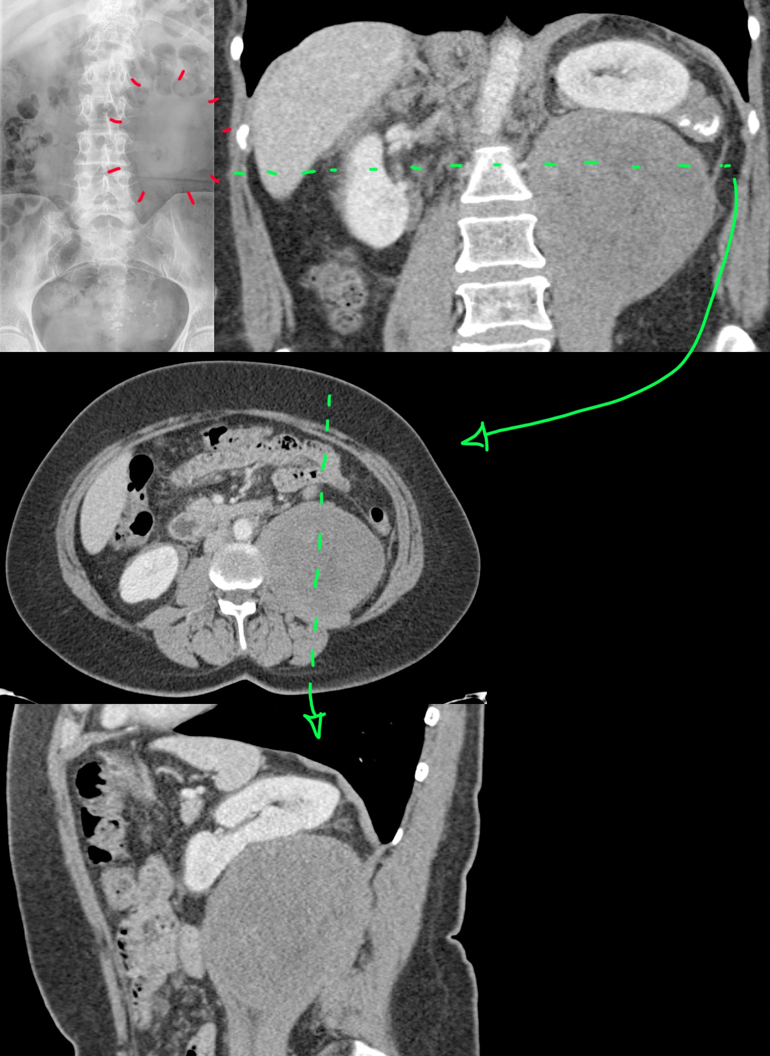
54 year old female with a history of uterine fibroids with persistent abdominal distention despite being told her fibroids shrunk after menopause - presumably via some pelvic ultrasound, which we didn't have.
Abdominal X-ray [top left] was possibly was called normal, which is not completely unreasonable. However, with the help of the follow-up coronal CT [top right], one can see that there is a vague area lacking any bowel gas that looks like it could be a mass. Axial [middle] and sagittal [bottom] CTs show a nonspecific, solid mass, well-circumscribed, with no calcifications, cysts, hemorrhage, or fat inside. The left kidney is pushed up by the mass, but the fat plane between the two is preserved with no evidence of renal invasion.
The patient underwent surgical resection.
Final diagnosis: Retroperitoneal dedifferentiated liposarcoma.
Tuberculous pancreatitis and lymphadenitis.

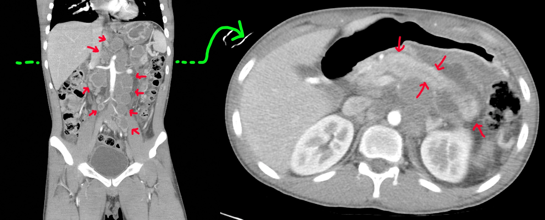
20 year old with a history of trisomy 21 and recent travel to Vietnam. Was seen there for nausea, vomiting, and abdominal pain with diagnosis of acute pancreatitis and what looked like lymphoma (but negative biopsy).
Coronal CT [left]: Big retroperitoneal lymph nodes with central cystic change/necrosis (red arrows).
Axial CT [right]: Done at the level of the green line shows a sausage-shaped pancreas compatible with ongoing versus resolving pancreatitis (red arrows).
Repeat EGD/EUS with celiac lymph node biopsy shows caseating granulomatous inflammation with positive culture and PCR for tuberculosis.
Bilateral indirect inguinal hernias. Appendix enters right inguinal hernia (Amyand hernia).

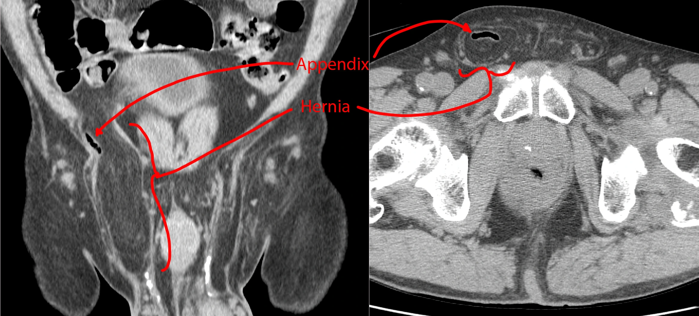
A companion case to this one. Bilateral groins with fat-containing hernias. The right-sided hernia also has a small air-filled tubular structure consistent with an appendix. There are no inflammatory changes to suggest acute appendicitis, so this is an incidental finding.
A right inguinal hernia containing an appendix is called an Amyand hernia and apparently pretty rare at <1% of inguinal hernias.
Bilateral indirect inguinal hernias. Ventriculoperitoneal shunt terminates in right inguinal hernia.

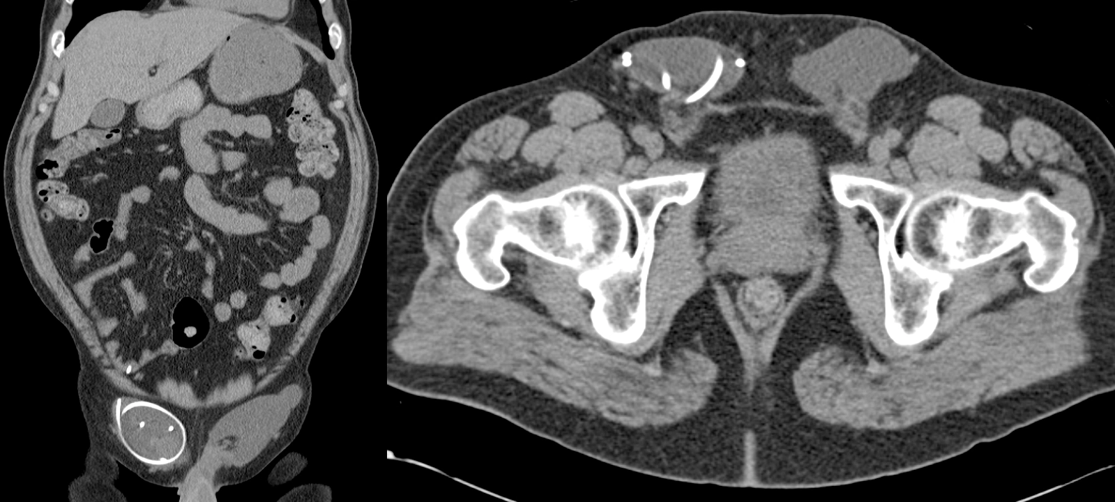
45 year old with a ventriculoperitoneal shunt (VPS) for history of toxoplasmosis. Incidental finding - no symptoms.
CT shows bilateral groin fluid collections. The right fluid collection contains the distal loops and tip of the VPS. So, just an incidental bit of cerebrospinal fluid in the groin.
Gastric obstruction from bezoar.

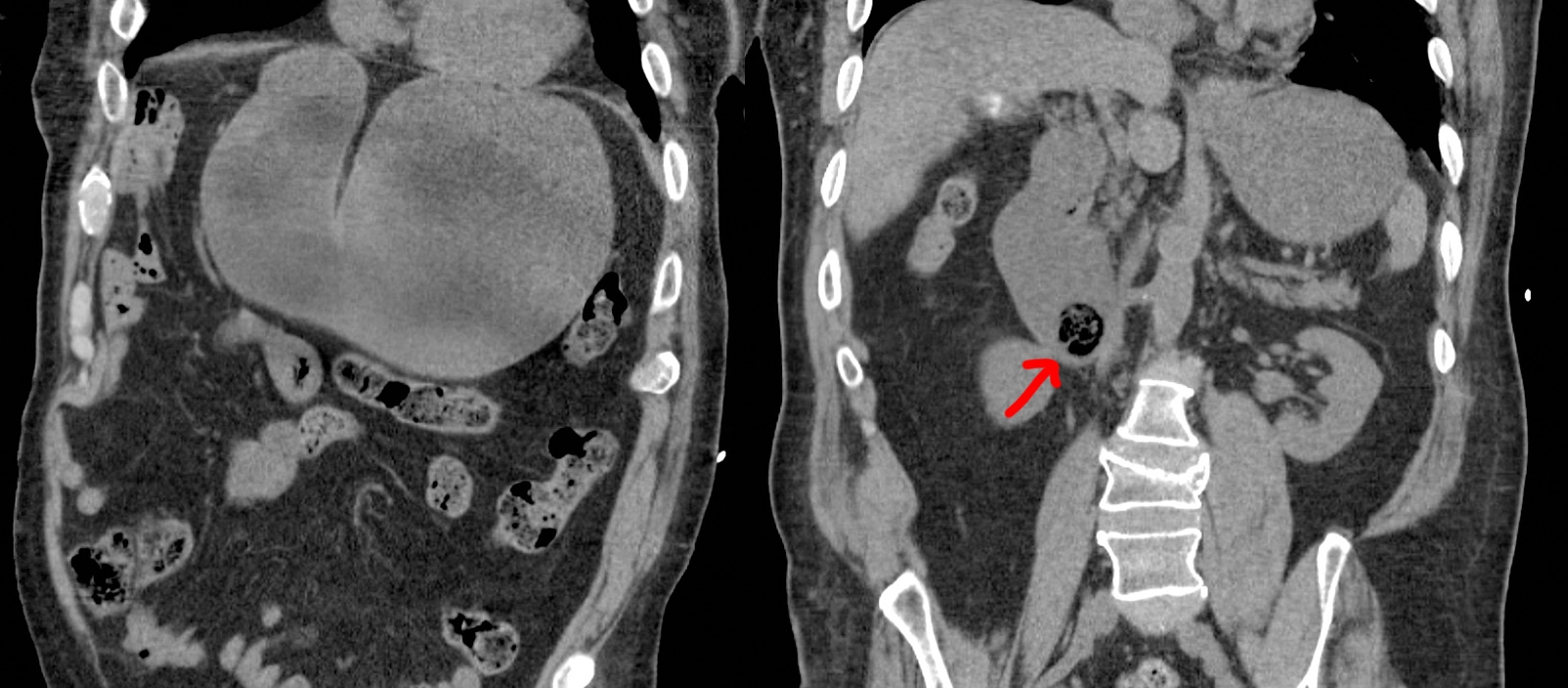
This patient with a history of developmental delay had a dental extraction 5 days before, during which they packed the area with gauze. The patient swallowed the gauze later on and started having nausea and vomiting 1 day before coming into the emergency department.
Coronal CT images show a distended stomach and a round object in the duodenum, representing a wad of gauze, as the source of the obstruction.